
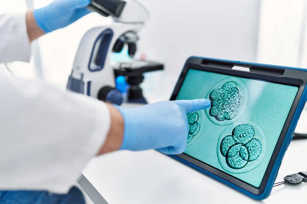
Determining the quality of an embryo and how ‘good’ it is involves reviewing the shape, structure and appearance to assess the potential for implantation.
Traditionally, embryologists monitored development by observing the embryo under a microscope each day, evaluating features such as cell division and tracking developmental changes throughout the culture process.
Recent technological advances, in the form of time-lapse imaging, now enable development to be monitored without the need to remove the embryos from the incubator.
Embryos are assessed and graded to identify those developing normally with the greatest potential to result in a successful pregnancy.
During the first three days of culture, embryo selection is typically based on several indicators of quality, including cell number, the rate of cell division, the uniformity of the cells, and the presence of cellular fragmentation. Embryologists pay close attention to the pace of embryo development, as irregular growth patterns may be associated with chromosomal abnormalities.
By the blastocyst stage (days 5–6), the embryo has enlarged significantly and developed into two distinct cell types. Quality is then assessed using a grading system consisting of a numerical score and two letter grades that evaluate the different parts of the embryo.
The numerical score (1–6) indicates the degree of blastocyst expansion, while the letter grades reflect the quality of the two cell types: the inner cell mass and the trophectoderm. Following implantation, the inner cell mass forms the foetus, whereas the trophectoderm gives rise to the placenta. Each structure is graded by the embryologist on a scale ranging from A (highest quality) to D (lowest quality).
The highest-quality available embryos are then selected for transfer.